Sample Preparation
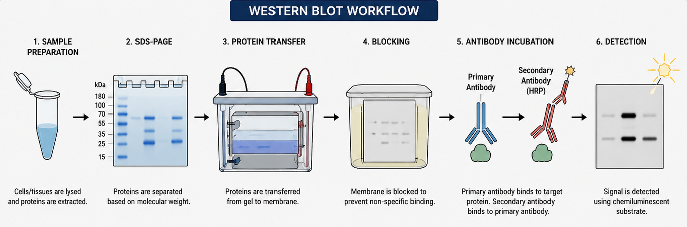
Proteins are extracted from Cells or tissues using lysis buffers such as RIPA or NP-40. A protein extraction buffer containing detergents and protease inhibitors to prevent degradation and preserve Post Translational Modifications.
Electrophoretic Separation (SDS-PAGE)
Proteins are separated by SDS-PAGE based on their molecular weight. The SDS provides a net-negative charge to the proteins, allowing them to migrate through a sieve-like gel matrix toward a positive electrode.
Protein Transfer
Separated proteins are transferred from the gel onto an adsorbent membrane (PVDF or nitrocellulose membrane) using electric current called as electroblotting. An important step to preserves the protein pattern from the gel and making proteins accessible for antibody based detection.
Blocking
To prevent non-specific antibody binding, the membrane (PVDF or nitrocellulose membrane) is "blocked" with reagents like non-fat dry milk or bovine serum albumin (BSA)
Antibody Incubation
The membrane is incubated with a primary antibody specific to the target protein. A secondary antibody labeled with enzymes like HRP (horseradish peroxidase) or fluorescent tag binds to the primary antibody. This enables detection and Visualization of the targeted protein
Detection
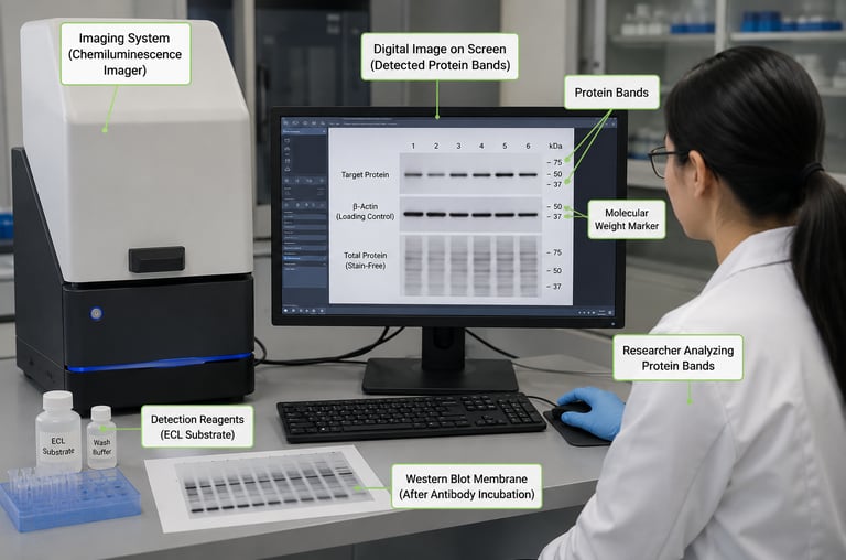
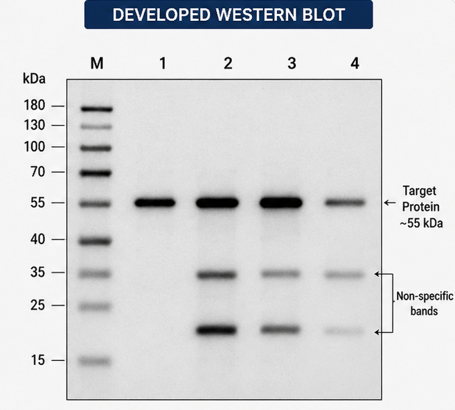
Protein bands are visualized using chemiluminescence or fluorescence detection systems. It is captured on X-ray film or digital imaging systems to quantify the protein's abundance.